Onion Skin Cell Microscope
Cells cell iodine plant 10x mount onion skin epidermis bulb light inner bubbles air animal nucleus eukaryotic microscopy structure fresh Onion cells microscope hi-res stock photography and images Onion skin 200x plant slides labelled dissection
Onion cells microscope hi-res stock photography and images - Alamy
Microscope cells microscopio cellula vegetale mm horizontal cellule zwiebel mikroskop zelle cipolla zellteilung Onion microscope structure staining microscopic schoolworkhelper biology shapes Microscopes – let’s go to the lab! 5°ep
Magnified 40x times 100x microscopy
Microscope onion under cellsOnion skin 10x epidermis mag inner not bulb nuclei obj staining phantom colours fig were microscopy The inner epidermis of the onion bulb’s cataphylls (the onion skin).Onion microscope cell stock epithelium under alamy.
Onion cells under microscope 40xOnion microscope cells under epidermal alamy epidermis light stock allium cepa high Onion skin microscope « optics & binocularsOnion cell cells microscope micrograph under 40x labeled stock alamy microscopic skin magnification root tip allium high epidermis bulb section.

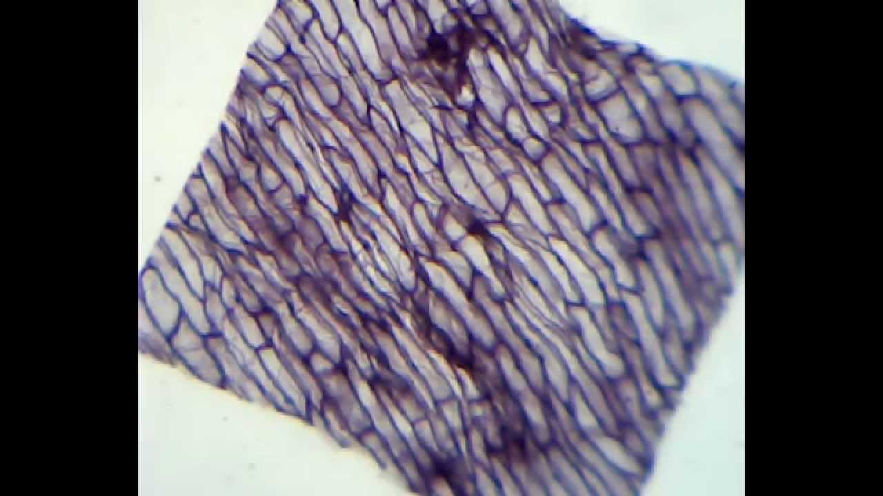
Onion cells microscope blue methylene stained under observation umberto flickr
Onion cells under microscopeOnion skin cells under microscope submited images. Onion microscope under 40x cells 100x 400xOnion cells under microscope.
The inner epidermis of the onion bulb’s cataphylls (the onion skin).Magnified microscope cell 40x microscopy micrographs walls Onion cell microscope hi-res stock photography and imagesOnion skin 200x.

Onion microscope 40x homage
The virtual microscopeOnion cells under the microscope: 40x Onion microscope skin cells eosin under cell stain vacuole epidermal stained cytoplasm mag look microscopy botany x40 why central thinOnion 400x cells microscopy forum think please any leave comments.
Onion cells under a microscopePlant & animal cells staining lab answers Microscope virtual 40x onion cell magnification next skinOnion skin cells under the microscope, horizontal field of view is.

Cells onion microscope under skin cell lab 100x lesson algae fungi weebly school light similarities eukaryotes differences prokaryotes magnification plant
Onion cell microscope hi-res stock photography and imagesOnion cells under microscope Onion cells at 400x.
.

ONION CELLS AT 400X - MicrobeHunter.com Microscopy Forum

Onion cells microscope hi-res stock photography and images - Alamy

Plant & Animal Cells Staining Lab Answers - SchoolWorkHelper

The Virtual Microscope

Onion skin cells under the microscope, horizontal field of view is

onion cells under microscope - YouTube

Onion Cells under Microscope

Onion Cells Under Microscope 40x